Ultraviolet Magnetic Circular Dichroism PhotoElectron Emission Microscopy
(UV MCD PEEM)
In 2006, we discovered substantial enhancements of magnetic circular dichroism
(MCD) near the work function threshold [see T. Nakagawa and T. Yokoyama,
Phys. Rev. Lett. 96 (2006) 237402]. In Ni(12monolayer)/Cu(001) we observed 100-1000 times
enhancement of the MCD signal compared to standard MOKE measurements. Using
the effect, we have been exploiting ultraviolet magnetic circular dichroism
photoelectron emission microscopy (UV MCD PEEM) with a femtosecond laser.
There have been no report on UV MCD PEEM of ultrathin films.
At present, we use as a light source a mode-locked Ti:sapphire laser (Spectra Physics, MaiTaiHP, 700-1000nm, 2.5W, 80MHz, <100fs) and higher-order harmonics generators (2nd, 3rd, 4th order), and the UV lights down to 210 nm are available almost continuously. On the other hand, as a PEEM apparatus, we have changed the apparatus from Elmitec PEEMSpector (spatial resolution 35 nm) to Omicron Focus IS-PEEM (spatial resolution 20 nm).
The image figure shows the UV MCD PEEM image from Cs-coated Ni(12monolayer)/Cu(001), where upward and downward magnetic domains are clearly distinguished. This is the first observation of UV MCD PEEM measurements for ultrathin films. Moreover, we have succeeded in the exploitation of two-photon photoemission MCD PEEM and femtosecond time resolving MCD PEEM systems.
See for details the following references.
T. Nakagawa and T. Yokoyama, Phys. Rev. Lett. 96 (2006) 237402 (4 pages).
T. Nakagawa, T. Yokoyama, M. Hosaka and M. Katoh, Rev. Sci. Instrum. 78 (2007) 023907 (5 pages).
T. Nakagawa, I. Yamamoto, Y. Takagi, K. Watanabe, Y. Matsumoto and T. Yokoyama,
Phys. Rev. B 79 (2009) 172404 (4 pages).
T. Nakagawa, K. Watanabe, Y. Matsumoto and T. Yokoyama, J. Phys. Condens. Mater. J. Phys.: Condens. Matter 21 (2009) 314010.
T. Nakagawa, I. Yamamoto, Y. Takagi and T. Yokoyama, J. Electron Spectrsc. Relat. Phenom. 181 (2010) 164-167.
K. Hild, G. Schönhense, H. J. Elmers, T. Nakagawa, T. Yokoyama, K. Tarafder
and P. M. Oppeneer, Phys. Rev. B82 (2010) 195430 (11 pages).
T. Yokoyama, T. Nakagawa and Y. Takagi, Int. Rev. Phys. Chem. 27 (2008) 449-505.
|
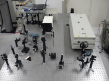 |
Photon energy tunable UV laser system |
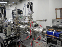 |
Photoelectron emission microscopy apparatus |
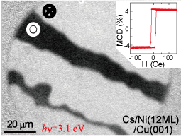 |
UV MCD PEEM image from Cs-coated Ni(12ML)/Cu(001) using a HeCd laser (325
nm). Dark and bright areas correspond to upward and downward magnetic domains,
respectively.
An inset gives a magnetization curve recorded separately. |
X-ray Magnetic Circular Dichroism (XMCD) using a superconducting magnet
X-ray magnetic circular dichroism (XMCD) is now widely applied to investigate
element specific magnetization and orbital magnetic moments. We have installed
a XMCD system with a superconducting magnet (JANIS, U.S.A.) at Beamline
4B (100-1000 eV, varied line spacing plane grating monochromator) of the
synchrotron radiation facility UVSOR-II. The measurement temperature is
5 K and the maximum magnetic field is 7 T (usually 5 T). The system is
equipped with a ultrahigh vacuum chamber for sample preparation, in which
sample preparation using Ar+ sputtering, heater, metal evaporation system
etc. and sample characterization using low energy electron diffraction
(LEED) and Auger electron spectroscopy apparatus can be done. Magnetic
thin films prepared in this chamber can be investigated in situ. Many users
including European researchers have studied with this system. For details,
see the folloing references.
T. Nakagawa, Y. Takagi, Y. Matsumoto and T. Yokoyama, Jpn. J. Appl. Phys. 47 (2008) 2132-2136.
X. D. Ma, T. Nakagawa, Y. Takagi, M. Przybylski, F. M. Leibsle, and T.
Yokoyama, Phys. Rev. B 78 (2008) 104420 (9 pages).
Y. Takagi, K. Isami, I. Yamamoto, T. Nakagawa and T. Yokoyama, Phys. Rev. B81 (2010) 035422 (8 pages).
I. Yamamoto, T. Nakagawa, Y. Takagi and T. Yokoyama, Phys. Rev. B81 (2010) 214442 (7 pages).
T. Yokoyama, T. Nakagawa and Y. Takagi, Int. Rev. Phys. Chem. 27 (2008) 449-505.
|
|
 |
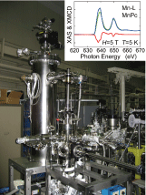
Ultrahigh vacuum superconducting magnet XMCD system. The inset gives an
XMCD example of Mn L-edge XMCD spectra of multilayer Mn phthalocyanine
taken at 5 K and 5 T.
|
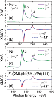
Fe and Ni L-edge XMCD spectra of Fe(2monolayer)/Ni(6monolayer) grown on
Pd(111). The angle theta is the X-ray incidence angle with respect to surface
normal. The X-ray direction is always parallel to the external magnetic
field.
|
Low-temperature Scanning Tunneling Microscopy (STM)
A scanning tunneling microscopic images can be taken at low temperature
at around 10 K. An Unisoku USM-1200 system is supplemented by a RHK Controller.
We use this system for the characterization of surface structures of magnetic
thin films etc. We have two similar systems, one of which was given by
Prof. Nishi after his retirement.
|
|
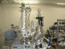
Unisoku low temperature STM apparatus. |
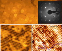 |
Examples of STM images. The sample is 2 monolayer gamma-Fe4N grown epitaxially
on Cu(001). An up-right photo gives the LEED pattern that exhibits p4gm(2x2)
superstructure. See for details, Takagi, K. Isami, I. Yamamoto, T. Nakagawa and T. Yokoyama, Phys. Rev. B81 (2010) 035422 (8 pages).
|
Magneto-Optical Kerr Effect (MOKE)
We can measure magnetization M-H curves of ultrathin magnetic thin films during sample preparation. The magnetic field is 3000 Oe and the sample temperature is 100-300 K using liq. Nitrogen. A photoelectron yield can also be detected. Several lasers as diodes 635nm, 405nm and 375nm, a HeCd layser (325nm) and a Ti:sapphire laser are available.
|
 |
 |
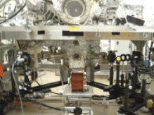
A MOKE system. Samples are prepared in a preparation chamber upward.
|
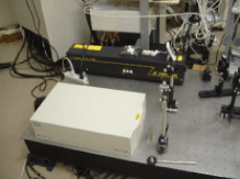
A Ti:sapphire laser MaiTai (Spectra Physics., 800nm, 100fs, 80MHz, 1W, white box) and a HeCdlaser (325nm, CW, 10mW ).
|
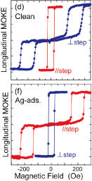 |
MOExamples of MOKE. The sample is a Co(5monolayer) film grown on a Cu(1
1 17) stepped surface. As Ag is deposited on the film, the magnetic easy
axis varies by 90 degrees from the step parallel direction to the step
perpendicular within the film plane. See for details, T. Nakagawa, H. Watanabe and T. Yokoyama, Phys. Rev. B74 (2006) 134422 (6 pages).
|


